UNIT # 2
CELLS, TISSUES, ORGANS AND ORGAN SYSTEMS
Day 1 THEME PAGE
Dec. 6th
The students were asked to look through the unit in the textbook (pages 2-99) and to find topics that they anticipate will be discussed in this unit. They are to use these ideas to create a theme page that includes the following criteria:
The students were asked to look through the unit in the textbook (pages 2-99) and to find topics that they anticipate will be discussed in this unit. They are to use these ideas to create a theme page that includes the following criteria:
| title_page_criterion.docx |
Due: Tuesday, December 13th
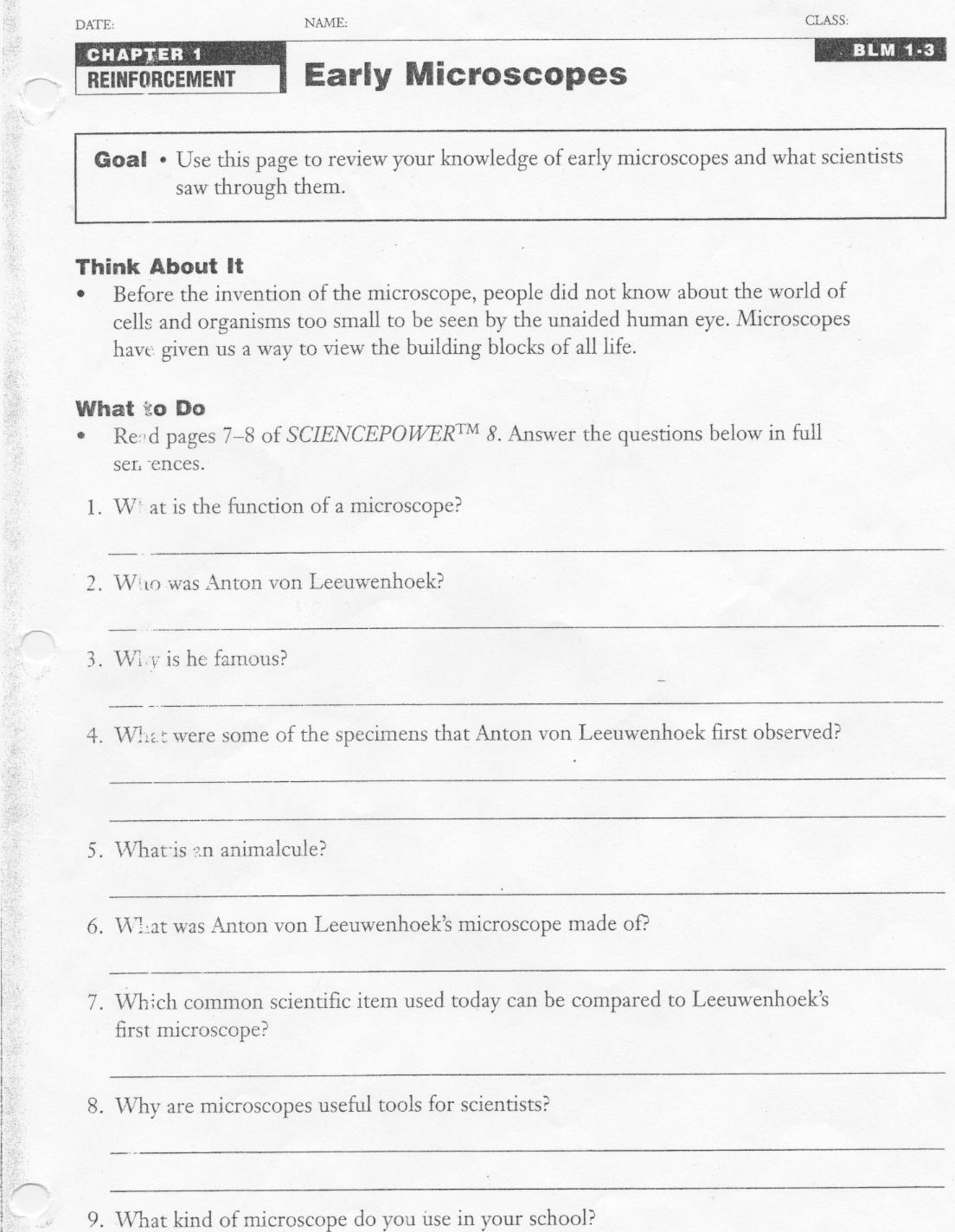
Day 2 THE EARLY MICROSCOPE
Dec. 7th
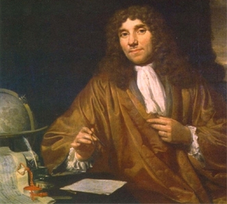
"The father of microscopy, Anton Van Leeuwenhoek of Holland (1632-1723), started as an apprentice in a dry goods store where magnifying glasses were used to count the threads in cloth. Anton van Leeuwenhoek was inspired by the glasses used by drapers to inspect the quality of cloth. He taught himself new methods for grinding and polishing tiny lenses of great curvature which gave magnifications up to 270x diameters, the finest known at that time.
These lenses led to the building of Anton Van Leeuwenhoek's microscopes (for more on the early microscope check out this weblink: http://bit.ly/9RbQtD)considered the first practical microscopes, and the biological discoveries for which he is famous. Anton Van Leeuwenhoek was the first to see and describe bacteria (1674), yeast plants, the teeming life in a drop of water, and the circulation of blood corpuscles in capillaries. During a long life he used his lenses to make pioneer studies on an extraordinary variety of things, both living and non-living, and reported his findings in over a hundred letters to the Royal Society of England and the French Academy." http://inventors.about.com/library/inventors/blleeuwenhoek.htm
The following clips provide a summary of early microscopy and those who were influential in advancing it. The first focusses on Von Leeuwenhoek and the second on Robert Hooke:
|
|
|
The Cell Theory Song recounts the components of the Cell theory and the major scientists that contributed to it.
Students were asked to complete the "Early Microscopes" activity sheet.
Due: Friday, December 9th
Due: Friday, December 9th
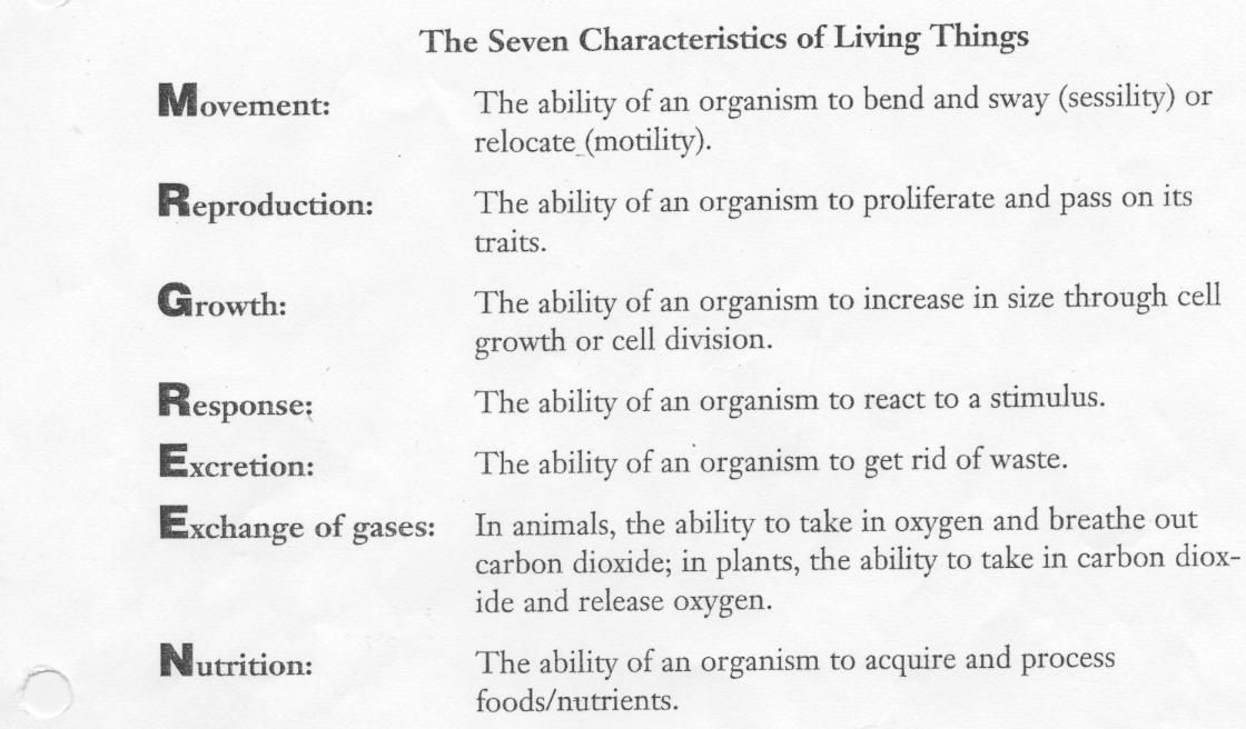
| 7_characteristics_of_living_things.jpg |
| early_microscopes.jpg |
Day 3 MICROSCOPES TODAY
Dec. 9th
Microscopes have come a long way from being instruments just used to see small objects in the 18th century. The invention of the microscope proved to be a major breakthrough in scientific and medical history. It was possible to look into human blood and see what was going on inside. Doctors and researchers could identify different bacteria and start creating medicines to counter them. It opened up avenues never explored earlier. In short it proved to be a huge boon to us human beings.
Microscopes Today
Microscopes are available in many different types and models. You can get economy ones as well as expensive ones. The best microscopes are made of metal alloys and are long lasting. Microscopes can be used in almost every sphere of life. They are used in schools, hospitals and clinical laboratories. They can be used for metallurgical and industrial work as well.
Types Of Microscopes
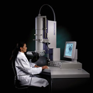
Different types of microscopes are used for different purposes. There are four types of biological microscopes that are used to examine tiny matter like blood cells and bacteria. These are the compound, dissection, transmission electron and scanning electron microscopes. Each one can be used for different functions. The compound microscopeallows you to view specimens at a high magnification power. It uses a fluorescent light to illuminate the specimens. The dissection microscope is like a regular compound microscope. It also uses light to illuminate the specimen. It has a lower magnification power and can be used to examine larger organisms by dissecting them. Electron microscopes use electrons instead of light to illuminate the specimens. These have very high magnifying power and resolution. They are used to examine blood and bacteria. They are especially useful in medical research. The digital microscopeis one of the latest innovations. It uses a USB to transfer images to a computer screen. This makes it easier to see the image and manipulate it on a larger screen. It also makes it easier to transmit such images to other scientists in other locations.
Microscopes are used in many locations now. The introduction to microscopes begins in school where they are used for educational purposes. Students conduct experiments by using dissection microscopes to examine small organisms. Microscopes are used in clinical and medical laboratories to examine blood samples to determine the kind of bacteria in them. This helps doctors diagnose their patients correctly and treat them accordingly. Microscopes are used extensively in research laboratories. Scientists are constantly examining different matter and specimens to learn more about them. Thanks to microscopes we discovered different diseases and the cures to many of them. Scientists are still conducting research by using microscopes to find cures and vaccines for many more life threatening diseases. There are microscopes used to examine non-biological specimens as well. They are used to examine and understand the composition of metals, rocks and even fossils. As you can see, microscopes are used in most areas of our lives.
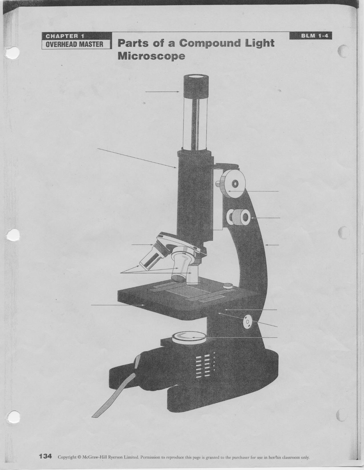
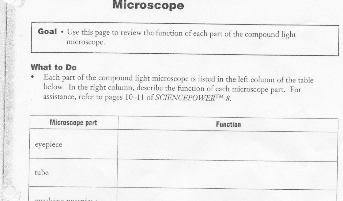
Today the students were asked to identify the location and function of each part of the compound light microscope. They were asked to complete the following activity pages.
The following weblink provides an (easy to read and) indepth explanation of the compound light microscope, how to use it properly and some prepared slides for examination. Enjoy!
http://bit.ly/rql3hZ
Microscopes Today
Microscopes are available in many different types and models. You can get economy ones as well as expensive ones. The best microscopes are made of metal alloys and are long lasting. Microscopes can be used in almost every sphere of life. They are used in schools, hospitals and clinical laboratories. They can be used for metallurgical and industrial work as well.
Types Of Microscopes
Different types of microscopes are used for different purposes. There are four types of biological microscopes that are used to examine tiny matter like blood cells and bacteria. These are the compound, dissection, transmission electron and scanning electron microscopes. Each one can be used for different functions. The compound microscopeallows you to view specimens at a high magnification power. It uses a fluorescent light to illuminate the specimens. The dissection microscope is like a regular compound microscope. It also uses light to illuminate the specimen. It has a lower magnification power and can be used to examine larger organisms by dissecting them. Electron microscopes use electrons instead of light to illuminate the specimens. These have very high magnifying power and resolution. They are used to examine blood and bacteria. They are especially useful in medical research. The digital microscopeis one of the latest innovations. It uses a USB to transfer images to a computer screen. This makes it easier to see the image and manipulate it on a larger screen. It also makes it easier to transmit such images to other scientists in other locations.
Microscopes are used in many locations now. The introduction to microscopes begins in school where they are used for educational purposes. Students conduct experiments by using dissection microscopes to examine small organisms. Microscopes are used in clinical and medical laboratories to examine blood samples to determine the kind of bacteria in them. This helps doctors diagnose their patients correctly and treat them accordingly. Microscopes are used extensively in research laboratories. Scientists are constantly examining different matter and specimens to learn more about them. Thanks to microscopes we discovered different diseases and the cures to many of them. Scientists are still conducting research by using microscopes to find cures and vaccines for many more life threatening diseases. There are microscopes used to examine non-biological specimens as well. They are used to examine and understand the composition of metals, rocks and even fossils. As you can see, microscopes are used in most areas of our lives.
Today the students were asked to identify the location and function of each part of the compound light microscope. They were asked to complete the following activity pages.
The following weblink provides an (easy to read and) indepth explanation of the compound light microscope, how to use it properly and some prepared slides for examination. Enjoy!
http://bit.ly/rql3hZ
Due: Dec. 13th
| the_parts_of_a_compound_light_microscope.jpg |
| the_compound_light_microscope.jpg |
The following clip outlines the locations of the parts of the compound light microscope and also how to use it safely.
THE ROAD TO DISCOVERY
The microscope, an invention that changed the way humans look at the world, came about through the process of much experimentation. Originating in the late 16th century, the microscope is certainly one histories most groundbreaking instruments.
The microscope opened up an entire new world to scientists and helped us to answer many questions about life.
In the first part of the lesson the students got the opportunity to research some of the original scientists who used microscopy to advance human knowledge of the world. The students were asked to complete the following under the title Road to Discovery using their Science Textbooks (pages 18-20) and information from the web:
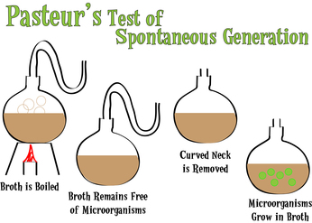
1) Explain the belief in "Spontaneous Generation." What did it help to answer for humans prior to the discovery of the microscope.
2) Complete "Check Your Understanding" questions page 20 #3-7.
3) Describe what the following scientists discovered using early microscope models:
a) Francesco Redi (1690)
b) Louis Pasteur (1864)
The microscope opened up an entire new world to scientists and helped us to answer many questions about life.
In the first part of the lesson the students got the opportunity to research some of the original scientists who used microscopy to advance human knowledge of the world. The students were asked to complete the following under the title Road to Discovery using their Science Textbooks (pages 18-20) and information from the web:
1) Explain the belief in "Spontaneous Generation." What did it help to answer for humans prior to the discovery of the microscope.
2) Complete "Check Your Understanding" questions page 20 #3-7.
3) Describe what the following scientists discovered using early microscope models:
a) Francesco Redi (1690)
b) Louis Pasteur (1864)
The following weblink provides an entertaining and informative review of the life of Louis Pasteur and his accomplishments through animation. They help to show the era that he was living in and the stresses he faced (including the deaths of 3 of his children to streptococcus disease) as he built up to his greatest discovery, the "Germ Theory" which revolutionized modern medicine.
http://www.toon.is/animated-hero-classics-louis-pasteur-video_5146111ff.html
http://www.toon.is/animated-hero-classics-louis-pasteur-video_5146111ff.html
The following clip provides an excellent description of the widely held belief in the theory"Spontaneous Generation" that people used to explain the appearance of animals like mice, frogs and flies. This clip goes on to show how this theory was later disproven by Francesco Redi in 1690.
UNICELLULAR ORGANISMS
A unicellular organism is any life form that consists of just a single cell. Most of life is unicellular, with bacteria serving as the majority. They are the oldest forms of life, and many existed 3.8 billion years ago, if not longer.
We can observe the larger unicellular organisms, such as amoebae, by using the higher settings on a light microscope. (Bacteria just appear as dots they are so tiny). To gather unicellular organisms for observation, you can place a cover slip on the surface of pond water, and leave it overnight. By the next morning, numerous unicellular organisms will have grown entire colonies on the bottom of the slip.
**Unicellular organisms replicate fast: colonies can double their size between 30 minutes and a few hours.
Some unicellular organisms have flagella (singular flagellum) which are little tails they use for locomotion. Others use many tiny hair like structures called cilia to move and eat, while still others use bloblike arms called pseudopods to get from one place to another to eat.
** The flagella of our unicellular ancestors is retained all the way up into the animals, where it makes an appearance in sperm. Source: http://www.wisegeek.com/what-is-a-unicellular-organism.htm
Today the students were asked to research a little about unicellular organisms, how they move and how they eat. They are to complete the following questions in their notebooks under the title, "Unicellular Organisms" They may use their Science textbooks and the web to help to complete the assignment.
1) Differentiate between the terms "unicellular" and "multicellular". What category (uni/multicellular) of organism do humans fit inot?
2) Where are most unicellular organisms found?
3) Differentiate between flagella and cilia.
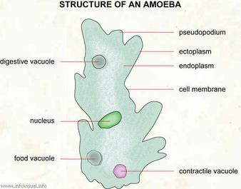
4) Describe (and sketch with labels) how the common unicellular organism call the Amoeba:
a) moves (the bloblike arms are called pseudopods) and
b) eats (called phagocytosis).
We can observe the larger unicellular organisms, such as amoebae, by using the higher settings on a light microscope. (Bacteria just appear as dots they are so tiny). To gather unicellular organisms for observation, you can place a cover slip on the surface of pond water, and leave it overnight. By the next morning, numerous unicellular organisms will have grown entire colonies on the bottom of the slip.
**Unicellular organisms replicate fast: colonies can double their size between 30 minutes and a few hours.
Some unicellular organisms have flagella (singular flagellum) which are little tails they use for locomotion. Others use many tiny hair like structures called cilia to move and eat, while still others use bloblike arms called pseudopods to get from one place to another to eat.
** The flagella of our unicellular ancestors is retained all the way up into the animals, where it makes an appearance in sperm. Source: http://www.wisegeek.com/what-is-a-unicellular-organism.htm
Today the students were asked to research a little about unicellular organisms, how they move and how they eat. They are to complete the following questions in their notebooks under the title, "Unicellular Organisms" They may use their Science textbooks and the web to help to complete the assignment.
1) Differentiate between the terms "unicellular" and "multicellular". What category (uni/multicellular) of organism do humans fit inot?
2) Where are most unicellular organisms found?
3) Differentiate between flagella and cilia.
4) Describe (and sketch with labels) how the common unicellular organism call the Amoeba:
a) moves (the bloblike arms are called pseudopods) and
b) eats (called phagocytosis).
The following clips may be useful when completing this assignment:
Day 7-8 PARTS OF PLANT AND ANIMAL CELLS
Jan.
Comparing Plant and Animal Cells
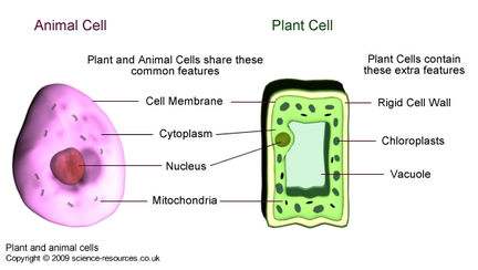
Multicellular organisms are differentiated from each other by being split into two categories based on the structure of the cells that they are composed of: Animal Cells and Plant Cells.
Organelles
Both plant and animal cells are composed of interior structures or parts that are enclosed within their own membrane inside of each cell. Each of these structures performs a particular function for the cell.
As you can see from the diagram above, plant and animal cells share a number of common organelles, however plant cells do have some extra features.
Today the students were asked to explore the following weblink called Biology4kids.com http://www.bam.gov/
Here they were asked to read through links (listed on right hand side of web page) that are associated with plant and animal cells and complete the accompaning quizzes (located at the bottom of each page):
1) Overview and Organelles
2) Cell Membranes
3) Cell Walls
4) Cytoplasm
5) Nucleus
6) Chloroplasts
7) Vacuoles
A virtual followup to this activity can be found at the following link: http://bit.ly/qBqo6n
As a followup, the students were asked to complete the following activity sheets:
Here are some additional video clips that can be viewed to help reinforce the similarities and differences of plant and animal cells.
PLANT AND ANIMAL CELL LAB
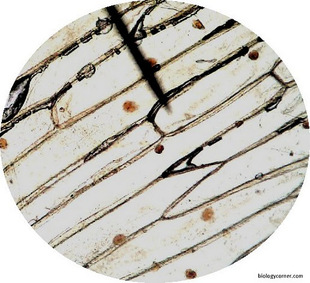
Onion Bulb Cell Stained with Iodine
High Power Maginification
To practice how to use a compound light microscope the students will be conducting a small group experiment in which they examine both plant and animal cells. They will have an opportunity to compare the similarities and differences between the two types of cells and will be required to write up a formal lab report (including all of the components of the scientific method). The students will be asked to create a hypothesis prior to the beginning of the lab. They will be required to prepare a plant cell slide (from an onion skin) and draw and label diagrams using different magifications. They will also draw and label diagrams of professionally prepared human skin cells. At the end of the lab, they will create conclusions based on their findings and answer analysis questions.
At Home Activity
The following video clip provides students with a chance to relive a cartoon classic from the animated series "The Magic School Bus." Many of them will be familiar with the show, but (hopefully) will have a better appreciation of the content of the show after having learned some of these concepts during this unit.
The instructions of how to prepare an onion skin cell are given in the following clip. There is also instructions concerning how to prepare a human skin cell using a swab on the inside of the mouth (gumline), however for sanitary reasons and a lack of time we simply use the professionally prepared human skin cells.
A fun recap of the parts of the cell that the students should be looking for can be found in the "Cell Cells - Parts of the Cell Rap" found in the clip below.
Note: For the purposes of our lab, the students will not be able to view chloroplasts because the onion bulb (from which we obtained our plant cells) grows underground and is thus not involved in photosynthesis.
The students are also not responsible for locating the mitchondria, the ribosomes or the endoplasmic reticulum that are mentionned in this rap. They will study these organelles in grade nine.
More At Home Fun
Try out this interactive game that helps to review ways to distinquish between plant and animal cells. It is called "The Mixed Up Cells". Professor Mill Ennium has left the Discovery Lab in the hands his trusty lab assistants. As one of his assistants, you find yourself exploring the lab for the first time on your own.
For Those Who Missed the Lab
A cool virtual biology lab can be found here: http://bit.ly/UhyCe6
A pre-lab showing the diagrams we will be creating in this lab can be found here and below that is a copy of the actual science lab report:
| onion_and_human_skin_pre-labs.docx |
A Closer Look at Cell Membranes
Diffusion of Molecules
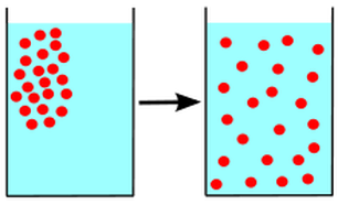
The cell membrane acts as a barrier between the inside and outside of the cell. There are two processes that allow for the transportation of molecules into and out of the cell (Active and Passive Transport). In grade eight, we concern ourselves only with passive transport (diffusion and osmosis). You will study Active Transport in grade nine.
Passive Transport involves the movement of molecules into and out of the cell without the cost of energy. In this case, the flow of molecules happens across a gradient. This means that the molecules will flow from an area of high concentration to an area of low concentration until an equal concentration is reached. In the diagram above, originally the molecules are closely packed together (high concentration), but over time they spread themselves out evenly.
Types of Membranes
Permeable - allowing substances to pass through easily (an open door)
Non-permeable - allowing no substances to pass through (a closed door)
Selectively Permeable - allowing certain substances, but not all substances to pass through (a screen on a door)
As the video says, humans have semi-permeable membranes.
Some molecules are able to pass into and out of the cell without any energy being used. Gases such as oxygen and carbon dioxide fit this category. When there is a high concentration of these gases on either side of the cell membrane, they will pass through the membrane until there is an equal concentration on both sides. This process is called Diffusion.
The diffusion of water molecules is so important to the human body that it is given it's own special name, Osmosis.
The following video shows an egg experiment that can be conducted in class that demonstrates the movement of water out of an egg (when placed in a sugary liquid) and the movement of water back into the wrinkled egg when it is resubmerged in pure water.
A Closer Look at the Nucleus - Mitosis (Cellular Reproduction)
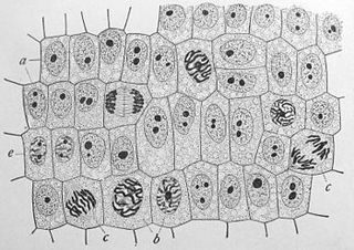
Plant Cells in Various Stages of Mitosis
The questions of why some people are taller than others; smarter than others; more musical than others; and more athletic than others, can be partially explained by the "genes" that make up our DNA codes. During grade eight, students are introduced (but not formally responsible for understanding) the topic of genetics.
Each individual cell in a human body contains the exact same genetic code. The process of a fetus starting at conception, made up of one individual cell, composed of a mixture of a mother's and a father's genes, eventually replicating, duplicating and diversifying itself to become a fully functioning child composed of trillions of cells, is truly one of life's greatest mysteries.
Today the students will examine a very basic introduction to the process of Mitosis. They will be responsible for briefly explaining and diagramming what happens to chomosomes during each of these stages of Prophase, Prometaphase, Metaphase, Anaphase and Telophase. Key vocabulary will include: chromosomes, spindle fibers, centromeres, poles, chromatid, mother and daughter cells
The following weblink provides a good basic description of the process of Mitosis and why it happens in eukaryotic cells. Eukaryotic refers to cells that contain membrane bound organelles. Plant and animal cells are eukaryotic. The website also provides some embedded words and video that should help you to familiarize students with the complicated process of DNA replication and duplication.
http://bit.ly/PKFQEi
neoK12 is an excellent website that provides interactive videos, lessons and games to reinforce and enhance the learning of mitosis. Check it out here: http://bit.ly/yWenHw
The following song is a fun way to help the students to remember the proper order of the five stages of Mitosis.
Cancer - "Mitosis Gone Wild"

The St. Mary Relay for Life is always a success
When asked, most students in my class can identify at least one person that they know who has suffered from cancer. Sadly, cancer is the number one cause of death in Canada. In 2012, an estimated 186,400 new cases of cancer (excluding non-melanoma skin cancer) were diagnosed in Canada and 75,700 cancer deaths occurred.
At St. Mary, we recognize the significance of these statistics and a fundraiser called "Relay for Life" is organized on our school grounds each spring. Students, teachers and parents form teams that take turns walking around a designated track lit up by covered candles inscribed with the names of loved ones who have suffered or died from cancer. The following weblink will take you to the "Canadian Cancer Society" webpage that shows how many teams participated last year at St. Mary and how much money was donated o cancer research from their efforts. A truly heartwarming event each year. http://bit.ly/OiCLPm
"Kids Health - What is Cancer?" is an excellent website for providing an introduction to the topic of cancer and what causes it (commonly referred to as a carcinogens). The website includes an audio setting that will allow students who struggle with reading to follow along as the highlighted text is read. This website also provides links to several other health related topics for parents, kids and teens with inquiring minds.
http://bit.ly/i3g0in
The website "Kids Against Cancer" provides a good list of common caricinogens on it's webpage entitled "The ABCs of Cancer."
You can find the website here: http://bit.ly/OQgkeD
The following provides a compilation of images and music (I Run for Life by Melissa Etheridge) in a inspiring video for those touched by breast cancer.
Introduction to Organ Systems in Humans
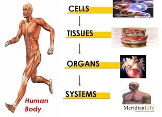
Levels of Organization in the Human Body
An adult human body is made up of 11 organ systems. These systems are composed of 22 internal organs, 600 muscles, 206 bones and 100 trillion cells.
Today students will define the words tissue, organ and organ system to help them to explain the levels of organization in the human body.
We will compare the levels of organization of a school system to the human body in order to help the students conceptualize how each of the levels becomes more complex. For example, the students would make up the largest component of the school system similar to the cells in the body. Teachers represent the second level of the school system and are compared to tissues. etc.
The following provides a basic definition of each of the levels of organization in the human body.
Level 1 - The Cell
A cell is the smallest functional and structural unit of all living organisms
Level 2 - Tissues
It is created by joining cells that have similar function or structure.
Level 3 - Organs
An organ is a group of tissues that perform a specific function or group of functions
Level 4 - Organ System
An organ system is a collection of organs that perform a specific function- the circulatory system or digestive system for example.
Level 5 - Organism
An organism is a being that's able to perform simple acts of survival. We humans are one for example.
The following website from Kids Health called "How the Body Works" is an excellent source of information for kids looking for an introduction to the study of tissues, organs and systems.
http://bit.ly/9D6ucr
The following video was produced by National Geographic. It tells the story of a human life, from first breath to last, told from within the body. It provides excellent detail about the cells, tissues, organs and organ systems. It pays particular attention to the importance of maintaining a healthy lifestyle to keep all of the systems functioning appropriately. Well worth the watch.
Body System #1 - The Digestive System
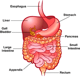
The human digestive system processes the food that we eat. It is made up of multiple organs and glands that digest the food, extract energy and nutrients, and later expel the waste byproducts.
The following weblink takes you to a very kid friendly tour of the digestive system. Perfect for the grade eight curriculum.
"Your Digestive System" by KidsHealth
http://bit.ly/xLLqYr
The website neoK12 provide interactive educational videos, lessons and games for school kids. A perfect site to get students engaged in researching the digestive system. Check it out here: http://bit.ly/s91TXc
Below is one of several excellent videos from the neoK12 website. Some of the videos are a little advanced for grade eight, but many are perfect for reinforcing learning.
As a followup to the lesson we watched the Bill Nye the Science Guy video entitled "Digestion" and completed an accompaning activity page.
Body System #2 - The Circulatory System
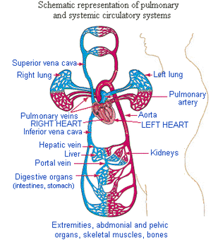
The human circulatory system consists of vessels, organs and muscles that help control the flow of blood and lymph around the body. It is collectively called the cardiovascular system and lymphatic system with the heart, arteries, capillaries, veins and blood being the main parts. The circulatory system moves the gases, nutrients and wastes too and from cells to maintain homeostasis. It helps fight diseases, maintains body temperature and pH.
The following weblink takes you to a very kid friendly tour of the circulatory system. Perfect for the grade eight
curriculum. "Your Circulatory System" by KidsHealth: http://bit.ly/SvAOKa
The website neoK12 provide interactive educational videos, lessons and games for school kids. A perfect site to get students engaged in researching more about the circulatory system. Check it out here: http://bit.ly/ikoLOy
As a followup to the lesson we watched two Bill Nye the Science Guy videos entitled "Blood and Circulation" and "The Human Heart" and completed the accompaning activity pages.
Body Systems #3 - The Respiratory System
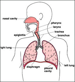
The respiratory system is made up of organs that help us to breathe. Our breathing supplies blood which is circulated through our body, allowing oxygen to reach all parts of the body. The respiratory system is composed of the mouth, nose, trachea, lungs and diaphragm. In the lungs, oxygen is absorbed while card
The following weblink takes you to a very kid friendly tour of the respiratory system. Perfect for the grade eight curriculum.
"Your Respiratory System" by KidsHealth: http://bit.ly/QDjvux
The website neoK12 provide interactive educational videos,
lessons and games for school kids. A perfect site to get students engaged in researching more about the respiratory system. Check it out here: http://bit.ly/QDjIxE
As a followup to the lesson we watched the Bill Nye the Science Guy video entitled "" and completed the
accompaning activity page.
Organ Systems and Vascular Tissue in Plants
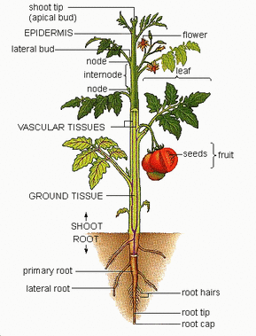
There are two organ systems in most plants:
1. The Root System (seeks and gathers water/nutrients from the soil)
2. The Shoot System (seeks light and adds height)
A third organ system is found in flowering plants:
The Reproductive System (female part = carpel, male part = stamen)
The website neoK12 provide interactive educational videos,
lessons and games for school kids. A perfect site to get students engaged in researching more about the plants. Check it out here: http://bit.ly/kJCeb4
Vascular Tissue
The vascular tissue in plants include xylem, which conducts water and minerals from the roots upward and throughout the plant, and phloem, which transports dissolved foods in all directions within the plant.
Transpiration
Transpiration is a process similar to evaporation. It is the loss of water from parts of plants (similar to sweating), especially in leaves but also in stems, flowers and roots.
A inclass demonstration of the transportation of liquid through a plants vascular tissue (fresh celery stalks) you can follow this weblink: http://bit.ly/btg4jK
The following video describes the movement of water (including transpiration) through a plant's xylem tissue:
If time permits, you may want to show the Bill Nye on Plants video. It contains a good overview of some of the issues discussed in the lesson (seed germination, photosynthesis etc). Unfortunately, it does not go into much about vascular tissue.
Unfortunately, I was unable to locate part III of the Bill Nye on Plants video. I will continue to search for it.
Unit Review
